(Study Material For DNL Students/ other Paramedical Students)
Understanding the Human Body
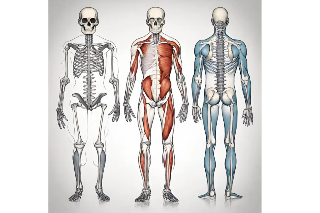
Human anatomy serves as the cornerstone of medical and health sciences, offering a profound insight into the intricate design and functioning of the human body. This discipline explores the structure of the body and how its various components work together to maintain life. Here, we will embark on an introductory journey into basic anatomy, covering the essential topics of the overview of human anatomy and the anatomical organization.
At its core, anatomy is about understanding the structure of organisms, and in the case of human anatomy, it focuses on the structures and systems that make up the human body.
To navigate this vast field, a grasp of basic anatomical terminology is indispensable. Terms such as anterior and posterior, proximal and distal, superior and inferior lay the foundation for precise communication within the anatomical community. These terms are crucial when describing the relative positions and relationships of body structures.

Anatomical planes such as sagittal, frontal, and transverse provide reference points for imaging and describing the body’s structures.
Directional terms, such as medial, lateral, superior, and inferior guide us in understanding the orientation of body parts.
Body cavities, including the cranial, abdominal, pelvic and thoracic cavities, house and protect vital organs, illustrating the intricate balance of form and function in anatomy.

Overview of the musculoskeletal system
Exploring the Musculoskeletal System:
The Dynamic Framework of the Human Body
The musculoskeletal system stands as the architectural framework of the human body, providing form, support, and the ability to move.
The musculoskeletal system comprising –muscles, bones, joints, and connective tissues
This intricate system orchestrates the marvel of human motion while maintaining structural integrity. In this part, we delve into the musculoskeletal setting, unraveling the dynamic interplay of components that define our physical capabilities.
Bone
At the core of the musculoskeletal system are the bones, serving as the rigid scaffolding upon which the entire body is built. Bones not only provide structural support but also protect vital organs, such as the heart and lungs. The skeletal system is a dynamic entity, constantly remodeling and adapting to the mechanical demands placed upon it. Through a process known as bone remodeling, old bone tissue is replaced by new, ensuring the maintenance of bone density and strength throughout life.
Joints
Joints, the articulations between bones, contribute to the remarkable flexibility and range of motion exhibited by the human body. Synovial joints, characterized by a synovial cavity filled with lubricating fluid, allow for smooth and controlled movement.
Different joint types including- hinge joints (e.g., elbow), ball-and-socket joints (e.g., shoulder and hip), and pivot joints (e.g., neck) facilitate a diverse array of movements, from simple flexion and extension to complex rotations.

Muscles
Connecting the bones and providing a dynamic interface for movement are the muscles. Skeletal muscles, attached to bones by tendons, contract and relax, generating the forces needed for movement. The arrangement of muscle fibers in parallel bundles imparts strength, while the precise coordination of muscle groups allows for intricate and controlled movements.
Muscles work in pairs, with agonist and antagonist muscles collaborating to produce coordinated actions and maintain joint stability.
Connective Tissue
An understanding of the musculoskeletal system is incomplete without acknowledging the intricate network of connective tissues that bind and support the various components. Ligaments, tough bands of fibrous tissue, connect bones to each other, providing stability to joints. Meanwhile, tendons connect muscles to bones, transmitting the forces generated during muscle contractions to produce movement. Fascia, a connective tissue network, envelops muscles and other structures, creating a seamless integration throughout the body.
Injuries
Injuries or disorders affecting the musculoskeletal system can significantly impact daily life. From fractures and sprains to conditions like osteoarthritis and rheumatoid arthritis, maintaining the health and functionality of the musculoskeletal system is paramount. Exercise, proper nutrition, and ergonomic practices contribute to the well-being of this dynamic system, promoting strength, flexibility, and longevity.
Basic Anatomy of the Spine
The spine, also known as the vertebral column or backbone, is a remarkable structure that serves as the central support for the human body. Its intricate design not only provides stability and protection but also allows for flexibility and movement. Understanding the anatomy of the spine is crucial for appreciating its role in maintaining posture, supporting bodily functions, and facilitating the complex interactions between the nervous system and the rest of the body.
Structure and Components:
The human spine is composed of 33 vertebrae, categorized into five regions: cervical (7), thoracic (12), lumbar (5), sacral (5 fused), and coccygeal (4 fused). Each vertebra is a distinct bone, and they are stacked upon each other, forming a flexible and curved column.

The vertebrae are separated by intervertebral discs, which act as shock absorbers and provide cushioning between the bony segments. These discs are made up of a tough outer layer called the annulus fibrosus and a gel-like inner core known as the nucleus pulposus. The intervertebral discs contribute to the spine’s flexibility while maintaining stability.
Vertebral Anatomy:
Each vertebra has specific features that contribute to its function within the spinal column. A typical vertebra consists of a vertebral body, a bony arch that encloses the spinal canal, and various processes extending from the arch. The vertebral foramen, formed by the vertebral body and the bony arch, houses the spinal cord, a crucial component of the central nervous system.

Processes include the spinous process, which projects backward and is palpable along the midline of the back, and transverse processes that extend laterally. Facet joints, located between adjacent vertebrae, allow for articulation and controlled movement. The vertebral arch, composed of the pedicles and laminae, surrounds and protects the spinal cord.
Spinal Cord and Nerves:
The spinal cord, a continuation of the brainstem, runs through the vertebral canal formed by the stacked vertebrae. It serves as the communication pathway between the brain and the peripheral nervous system.

Nerve roots, emerging from the spinal cord, pass through intervertebral foramina, connecting the spinal cord to the various organs and muscles throughout the body.
Supporting Structures:
Ligaments and muscles surrounding the spine play a crucial role in providing stability and facilitating movement. Ligaments, such as the anterior and posterior longitudinal ligaments, connect and support the vertebrae.

Muscles, arranged in layers along the spine, contribute to posture, movement, and protection.
Common Spinal Conditions:
Understanding spinal anatomy is integral to recognizing and managing various spinal conditions. Issues such as herniated discs, spinal stenosis, scoliosis, and degenerative disc disease can affect the spine’s structure and function.

Knowledge of spinal anatomy aids in diagnosis, treatment planning, and preventive measures for maintaining spinal health.
Basic Anatomy of the Thoracic Cage
The thoracic cage, an intricate bony framework, forms the foundation of the thoracic region, playing a pivotal role in protecting vital organs and supporting respiratory function. Comprising the sternum, ribs, and thoracic vertebrae, this anatomical structure is fundamental to the stability and functionality of the upper body.
At the anterior midline of the thoracic cage lies the sternum, a flat bone that connects to the ribs via costal cartilage. The sternum consists of three parts: the manubrium, body, and xiphoid process. The manubrium articulates with the clavicles and the first rib, contributing to the uppermost part of the thoracic cage. Connected to the sternum are twelve pairs of ribs, forming the lateral and posterior aspects of the thoracic cage. These ribs articulate with the thoracic vertebrae posteriorly and curve anteriorly, contributing to the dynamic structure of the ribcage.
The thoracic vertebrae, aligned along the posterior aspect of the thoracic cage, provide essential support and contribute to the overall flexibility of the spine. The articulation of the ribs with the thoracic vertebrae creates a protective enclosure around the thoracic cavity, housing vital organs such as the heart and lungs.
Respiratory function is a key aspect of the thoracic cage’s role. During inhalation, the ribcage expands as the ribs elevate, lifting the sternum and increasing the volume of the thoracic cavity. Simultaneously, the diaphragm contracts, enhancing the thoracic space for air intake into the lungs. Exhalation involves the relaxation of the ribcage and diaphragm, resulting in a reduction of thoracic volume and the expulsion of air from the lungs. This intricate dance of bones and muscles allows for the dynamic process of breathing, essential for sustaining life.
Understanding the basic anatomy of the thoracic cage provides a foundation for appreciating its multifaceted functions. From protecting vital organs to facilitating respiratory processes, the thoracic cage stands as a testament to the complexity and precision of the human body’s design.
Basic Anatomy of the Upper Limb
Now embarking on the journey of understanding human anatomy, delving into the intricacies of the upper limb unveils a fascinating world of interconnected structures and functions. Comprising the shoulder, arm, forearm, wrist, and hand, the upper limb is a marvel of biomechanical design.

At the core of the upper limb’s skeletal structure lies the shoulder girdle, connecting the limb to the axial skeleton. The humerus, the long bone of the arm, articulates with the scapula and clavicle, forming the foundation for a myriad of movements. Moving distally, the forearm showcases the dynamic interplay of the ulna and radius, contributing to the versatility of hand movements.
The wrist, a complex arrangement of eight carpal bones, serves as a junction between the forearm and hand. This intricate network facilitates both stability and a wide range of motions. Further down, the hand, adorned with metacarpals and phalanges, reveals the precision and dexterity required for daily activities.

As we explore the basic anatomy of the upper limb, we unlock the secrets of muscle attachments, joint articulations, and the neurological networks that orchestrate movement. This foundational knowledge lays the groundwork for future medical practitioners, therapists, and enthusiasts alike, fostering a profound appreciation for the beautifully engineered mechanics of the human upper limb.
Basic Anatomy of the Lower Limb
The lower limb, a marvel of biomechanical engineering, encompasses the regions of the hip, thigh, knee, leg, ankle, and foot. Anchored by the hip joint, the femur, which stands as the body’s longest bone, sets the stage for a cascade of interconnected skeletal components. Progressing through the thigh, the femur establishes crucial articulations with the tibia and fibula in the leg, forming an indispensable framework for weight-bearing activities.

Descending further, the anatomy of the ankle and foot unfolds a meticulous arrangement of tarsal, metatarsal, and phalangeal bones, constituting the foundation for stability and facilitating a spectrum of nuanced movements. Ligaments and tendons intricately interlace within this complex structure, ensuring the seamless integration of bones, muscles, and joints.

The comprehension of the basic anatomy of the lower limb holds paramount significance for professionals in healthcare, athletes, and enthusiasts alike. As we peel back the layers of muscle, bone, and connective tissue, profound insights into the biomechanical intricacies that propel us forward are gleaned. This foundational knowledge lays the groundwork for a comprehensive understanding of injuries, rehabilitation, and the symphonic interplay inherent in the human motion choreographed by the lower limb.